×


پیوند ها
آمار بازدید سایت
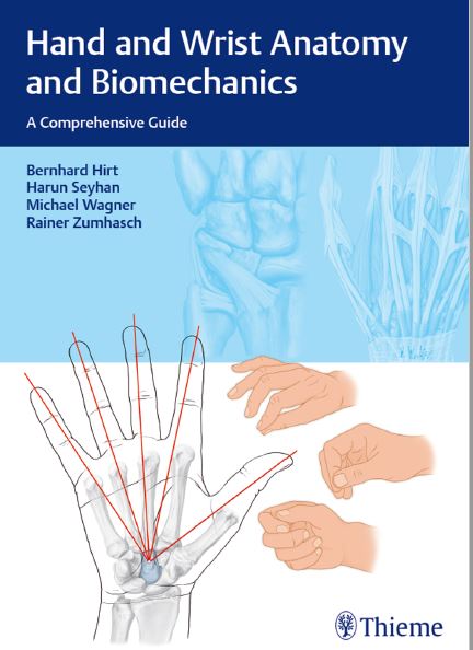
1 Anatomy and Functional Anatomy of the Hand . . . . . . . . . . . . . . . . . . . . . . . . . . . . . . . . . . . . . . . 2
1.1 Introduction . . . . . . . . . . . . . . . . . . . . . . . . . . 2
1.2 Structure and Function of the Proximal
and Distal Radioulnar Joints . . . . . . . . . . . . 2
1.2.1 Anular Ligament of the Radius . . . . . . . . . . . . . 3
1.2.2 Oblique Cord . . . . . . . . . . . . . . . . . . . . . . . . . . . 4
1.2.3 Interosseous Membrane of the
Forearm . . . . . . . . . . . . . . . . . . . . . . . . . . . . . . . 4
1.2.4 Structure and Function of the
Triangular Fibrocartilage Complex . . . . . . . . . 6
Ulnocarpal Disc and Meniscal Homologue . . . . . . 8
Palmar and Dorsal Radioulnar Ligaments . . . . . . . 8
Ulnolunate and Ulnotriquetral Ligaments . . . . . . . 8
Ulnar Collateral Ligament of Wrist Joint . . . . . . . . 8
Tendon Sheath of the Extensor Carpi Ulnaris
Muscle . . . . . . . . . . . . . . . . . . . . . . . . . . . . . . . . . 9
1.2.5 Muscles of the Radioulnar Joint—Pronation
and Supination . . . . . . . . . . . . . . . . . . . . . . . . . 9
Pronator Muscles . . . . . . . . . . . . . . . . . . . . . . . . . 9
Supinator Muscles . . . . . . . . . . . . . . . . . . . . . . . 10
1.3 Wrist and Movement Axes . . . . . . . . . . . . 10
1.3.1 Movement Axes . . . . . . . . . . . . . . . . . . . . . . . . 10
1.3.2 Structure and Function of theWrist
Joint . . . . . . . . . . . . . . . . . . . . . . . . . . . . . . . . . 12
Carpal Bones . . . . . . . . . . . . . . . . . . . . . . . . . . . 12
Joints . . . . . . . . . . . . . . . . . . . . . . . . . . . . . . . . . 14
1.3.3 Ligament System andWrist Stability . . . . . . 16
Superficial Layer of the Ligaments of the Wrist 17
Middle Layer of Ligaments of the Hand . . . . . . . 19
Deep Layer of the Ligaments of the Hand . . . . . 23
Conclusions . . . . . . . . . . . . . . . . . . . . . . . . . . . . 25
1.3.4 Muscles of theWrist: Extension–Flexion,
Radial Deviation–Ulnar Deviation, and
Circumduction . . . . . . . . . . . . . . . . . . . . . . . . 25
Muscles Involved in Flexion and Ulnar Deviation
of the Wrist . . . . . . . . . . . . . . . . . . . . . . . . . . . . 26
Muscles Involved in Extension and Radial
Deviation of the Wrist . . . . . . . . . . . . . . . . . . . . 28
1.3.5 Arthrokinematics of theWrist Joint . . . . . . . 28
Kinematics of the Wrist: General Aspects . . . . . . 30
Arthrokinematics of the Wrist Joint: Specific
Aspects . . . . . . . . . . . . . . . . . . . . . . . . . . . . . . . 30
1.3.6 Other Important Anatomical Structures
of theWrist . . . . . . . . . . . . . . . . . . . . . . . . . . . 33
Dorsal and Palmar Tendon Compartments . . . . 33
Carpal Tunnel, Ulnar Tunnel, and Innervation
of the Hand . . . . . . . . . . . . . . . . . . . . . . . . . . . . 38
Blood Supply to the Wrist Joint and Hand . . . . . 46
Lymphatics of the Hand . . . . . . . . . . . . . . . . . . . 47
1.4 Thumb . . . . . . . . . . . . . . . . . . . . . . . . . . . . . . 48
1.4.1 Movements and Range of Motion . . . . . . . . . 48
Movements of the Thumb CMC Joint . . . . . . . . . 48
Range of Motion of the Thumb MCP and
IP Joints . . . . . . . . . . . . . . . . . . . . . . . . . . . . . . . 49
1.4.2 Structure and Function of the Thumb
CMC Joint . . . . . . . . . . . . . . . . . . . . . . . . . . . . . 49
Ligaments of the Thumb CMC Joint . . . . . . . . . . 51
Muscles of the Thumb CMC Joint . . . . . . . . . . . . 54
1.4.3 Structure and Function of the Thumb
MCP and IP Joints . . . . . . . . . . . . . . . . . . . . . . 57
MCP Joint of the Thumb . . . . . . . . . . . . . . . . . . 58
IP Joint of the Thumb . . . . . . . . . . . . . . . . . . . . . 59
1.5 Structure and Function of the Palm . . . . 60
1.5.1 Structure and Function of the Second to
Fifth Metacarpals . . . . . . . . . . . . . . . . . . . . . . 60
1.5.2 Muscles of the Metacarpals . . . . . . . . . . . . . . 62
Dorsal and Palmar Interossei . . . . . . . . . . . . . . . 62
Lumbricals . . . . . . . . . . . . . . . . . . . . . . . . . . . . . 63
Abductor Digiti Minimi Muscle . . . . . . . . . . . . . 63
Flexor Digiti Minimi Brevis Muscle . . . . . . . . . . . 64
Opponens Digiti Minimi Muscle . . . . . . . . . . . . . 64
1.5.3 Palmar Aponeurosis (Metacarpal Area) . . . . 64
Longitudinal Fibers . . . . . . . . . . . . . . . . . . . . . . 65
Transverse Fibers . . . . . . . . . . . . . . . . . . . . . . . . 66
1.6 Structure and Function of the Finger
Joints . . . . . . . . . . . . . . . . . . . . . . . . . . . . . . . . 66
1.6.1 Structure and Function of the
MCP Joints . . . . . . . . . . . . . . . . . . . . . . . . . . . . 66
Collateral Ligaments . . . . . . . . . . . . . . . . . . . . . 67
Palmar and Dorsal Connective Tissue Plate . . . . 68
Deep and Superficial Transverse Metacarpal
Ligaments . . . . . . . . . . . . . . . . . . . . . . . . . . . . . 69
1.6.2 Structure and Function of the
PIP Joints . . . . . . . . . . . . . . . . . . . . . . . . . . . . . 69
Collateral Ligaments of the PIP Joint . . . . . . . . . 70
Palmar and Dorsal Fibrocartilage Plate . . . . . . . . 70
Anular and Cruciate Ligaments . . . . . . . . . . . . . 70
Dorsal Digital Expansion . . . . . . . . . . . . . . . . . . 72
1.6.3 Structure and Function of the
DIP Joints . . . . . . . . . . . . . . . . . . . . . . . . . . . . . 73
1.6.4 Range of Motion of the MCP, PIP, and
DIP Joints . . . . . . . . . . . . . . . . . . . . . . . . . . . . . 75
▶ Anatomical structure of the carpal tunnel. The carpal
tunnel is an osteofibrous canal, approximately 2.5cm
long, on the palmar side of the hand (▶Fig. 1.40). In the
proximal section, the floor is formed by the capitate,
hamate, and triquetrum and in the distal section by the
capitate and trapezoid. The radial wall is formed by the
proximal scaphoid and the distal trapezium, and the
ulnar wall by the proximal triquetrum and distal hamate.
The roof of the carpal tunnel is formed by the flexor retinaculum,
which inserts onto the radial aspect at the scaphoid
tubercle and the tubercle of the trapezium and on
the ulnar aspect, at the pisiform and hamate tubercle. The
flexor retinaculum is thicker in the distal portion and the
carpal space narrows in this less superficial location.27
Two palmar tendon sheaths traverse the carpal tunnel, in
the second palmar tendon compartment the flexor pollicis
longus muscle, and in the third palmar tendon compartment
the eight tendons of the flexor digitorum
profundus and superficialis muscles. The median nerve
runs between the third tendon compartment and the
flexor retinaculum; its course is very variable, but usually
radial of the middle line.27
برچسب های مهم
 فروش فیلتر بورسی استریکلی فقط 75 هزار تومان
 کسب درآمد اینترنتی 300000 تومان در خانه در کمتر از 30 دقیقه
 کسب و کار اینترنتی با درآمد میلیونی
 روش درآمدزایی در خواب (تعجب نکنید! کلیک کنید و بخوانید)
 ربات همه کاره اینستاگرام
 کسب درآمد از هوش مصنوعی ماهیانه حداقل ۱۲ میلیون تضمینی
 کسب درآمد روزانه حداقل یک میلیون تومان ! کاملا حلال و واقعـی !!
 کسب درآمد ابدی و بی نهایت 100% واقعی
 مجموعه ی آموزش تعمیر لامپ کم مصرف (از مبتدی تا پیشرفته)
 افزایش ممبر کانال، گروه و ربات تلگرام به صورت بی نهایت (اد ممبر)
 دانلود مجموعه آموزشی پایپینگ ( Piping ) و نقشه خوانی + آموزش سه نرم افزار طراحی و تحلیل لوله کشی صنعتی
 آموزش برنامه نویسی آردوینو
 آموزش بازكردن انواع قفل ها بدون كليد(ويژه)
 آموزش رایگان کسب درآمد از سایت الیمپ ترید ( olymp trade )
 کسب و کار اینترنتی در منزل
 بازگردانی پیامک های حذف شده- ریکاوری پیامک ۱۰۰٪ عملی
 اموزش ویرایش امضا و پکیج برنامه اندروید و کسب درامد از مارکت های اندرویدی
 آموزش ساخت بازی بدون دانش برنامه نویسی و طراحی سه بعدی مبتدی تا پیشرفته با نرم افزار
 کد های آماده html و css جهت یادگیری و طراحی سریع
 اموزش کسب درامد از اینترنت روزانه ۳میلیون تومان تضمینی و تست شده 1403
 درامدزایی در خواب! (تعجب نکنید! بخوانید)
 نسخه خطی اشعار و پیشگویی های شاه نعمت الله ولی
 آموزش كامل تعمير لامپ كم مصرف(اختصاصي)
 نسخه خطی اشعار و پیشگویی های شاه نعمت الله ولی
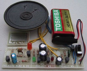 مدار داخلی واکی تاکی(اموزش ساخت)
 کتاب افزایش ممبر کانال تلگرام
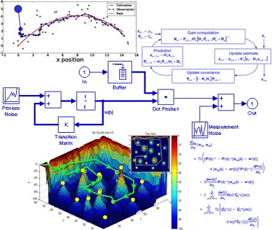 دانلود پاورپوینت فیلتر کالمن بر روی یک سنسور شتاب سنج برای تخمین سه متغیر حالت
 اندیکاتور ای کیو آپشن
 دانلود100% رایگان نرم افزار تبلیغات در تلگرام + آموزش کامل و فیلم آموزشی
moballegh1149@gmail.com


