×


پیوند ها
آمار بازدید سایت
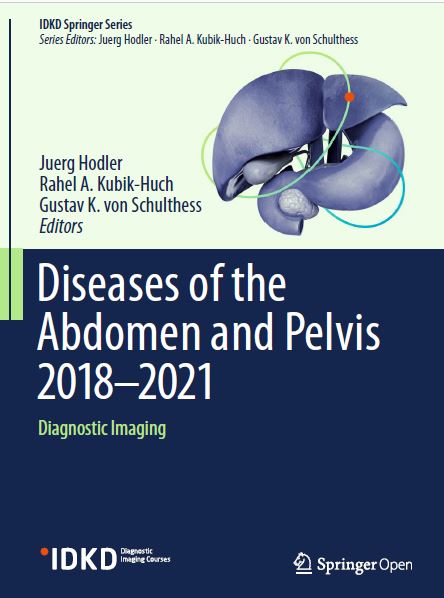
1 Renal Tumors........
Lejla Aganovic and Richard H. Cohan 2 MRI of the Pelvic Floor and MR Defecography ....
Francesca Maccioni and Celine D. Alt 3 Benign Disease of the Uterus.
Karen Kinkel, Susan M. Ascher, and Caroline Reinhold 4 Therapy Monitoring of Oncologic Disease in the
Abdomen (Including PET/CT)..
Irene A. Burger and Regina G. H. Beets-Tan 5 Disease of the Gallbladder and Biliary Tree.......
Jeong Min Lee and Daniel T. Boll 6 Pathways for the Spread of Disease in the Abdomen and Pelvis
James A. Brink and Brent J. Wagner 7 Urogenital Pathologies in Children Revisited.....
Jeanne S. Chow and Annemieke S. Littooij 8 Adnexal Diseases.
Andrea Rockall and Rosemarie Forstner
Adrenal Imaging..
Isaac R. Francis and William W. Mayo-Smith 10 Diseases of the Upper GI Tract
Richard M. Gore and Marc S. Levine 11 Magnetic Resonance Imaging of the Prostate
in the PI-RADS Era ... Bernd Hamm and Patrick Asbach
12 Small Bowel Disease......
Andrea Laghi and Amy K. Hara 13 Emergency Radiology of the Abdomen and Pelvis:
Imaging of the Non-traumatic and Traumatic Acute Abdomen.... Jay P. Heiken, Douglas S. Katz, and Yves Menu
14
15
Diseases of the Pancreas..... Thomas K. Helmberger and Riccardo Manfredi The Pediatric Gastrointestinal Tract: What Every Radiologist Needs to know. Emily A. Dunn, Øystein E. Olsen, and Thierry A. G. M. Huisman
16 Diseases of the Colon and Rectum: CT Colonography ........
C. Dan Johnson and Perry J. Pickhardt 17 Focal Liver Lesions....
Wolfgang Schima, Dow-Mu Koh, and Richard Baront 18 Malignant Diseases of the Uterus
Yulia Lakhman and Caroline Reinhold 19 Benign Diseases of the Colon and Rectum (incl. CT colonography).
Thomas Mang and Philippe Lefere 20 Urinary Obstruction, Stone Disease, and Infection...............
Refky Nicola and Christine O. Menias
21 Diffuse Liver Disease: Cirrhosis, Focal Lesions in Cirrhosis,
and Vascular Liver Disease.....
Khoschy Schawkat and Caecilia S. Reiner 22 Imaging of Diffuse and Inflammatory Liver Disease
Pablo R. Ros 23 Urinary Obstruction, Stone Disease, and Infection.............
S.O. Schönberg, J. Budjan, and D. Hausmann 24 Imaging of Testicular and Scrotal Masses: The Essentials
Andreas G. Wibmer and Hebert Alberto Vargas
gonadal vessels down into the pelvis, and the presence of
follicles or a corpus luteum cyst, if present, is confirmatory.
If two normal ovaries are identified, then the
AdnexMR score is 1, and the MRI should focus on characterizing
the non-ovarian mass.
2. If there is an ovarian lesion, then the next question is
whether the cyst has benign features. This could include
a unilocular cyst with no enhancing solid tissue, a typical
endometrioma, or a mature cystic teratoma. Lesions with
no wall enhancement or lesions with solid tissue that are
homogeneously low signal intensity on both T2 and high
b value DWI, with low enhancement, are almost certainly
benign. These lesions all fall into an AdnexMR
score of 2.
برچسب های مهم
 فروش فیلتر بورسی استریکلی فقط 75 هزار تومان
 کسب درآمد اینترنتی 300000 تومان در خانه در کمتر از 30 دقیقه
 کسب و کار اینترنتی با درآمد میلیونی
 روش درآمدزایی در خواب (تعجب نکنید! کلیک کنید و بخوانید)
 ربات همه کاره اینستاگرام
 کسب درآمد از هوش مصنوعی ماهیانه حداقل ۱۲ میلیون تضمینی
 کسب درآمد روزانه حداقل یک میلیون تومان ! کاملا حلال و واقعـی !!
 کسب درآمد ابدی و بی نهایت 100% واقعی
 مجموعه ی آموزش تعمیر لامپ کم مصرف (از مبتدی تا پیشرفته)
 افزایش ممبر کانال، گروه و ربات تلگرام به صورت بی نهایت (اد ممبر)
 دانلود مجموعه آموزشی پایپینگ ( Piping ) و نقشه خوانی + آموزش سه نرم افزار طراحی و تحلیل لوله کشی صنعتی
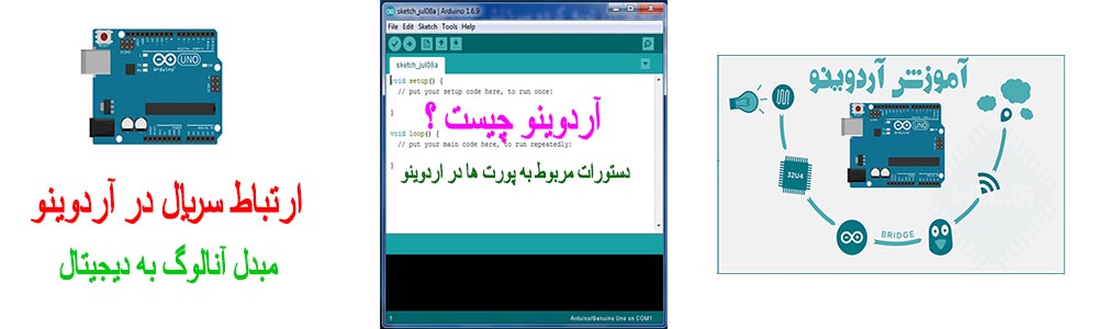 آموزش برنامه نویسی آردوینو
 آموزش بازكردن انواع قفل ها بدون كليد(ويژه)
 آموزش رایگان کسب درآمد از سایت الیمپ ترید ( olymp trade )
 کسب و کار اینترنتی در منزل
 بازگردانی پیامک های حذف شده- ریکاوری پیامک ۱۰۰٪ عملی
 اموزش ویرایش امضا و پکیج برنامه اندروید و کسب درامد از مارکت های اندرویدی
 آموزش ساخت بازی بدون دانش برنامه نویسی و طراحی سه بعدی مبتدی تا پیشرفته با نرم افزار
 کد های آماده html و css جهت یادگیری و طراحی سریع
 اموزش کسب درامد از اینترنت روزانه ۳میلیون تومان تضمینی و تست شده 1403
 درامدزایی در خواب! (تعجب نکنید! بخوانید)
 نسخه خطی اشعار و پیشگویی های شاه نعمت الله ولی
 آموزش كامل تعمير لامپ كم مصرف(اختصاصي)
 نسخه خطی اشعار و پیشگویی های شاه نعمت الله ولی
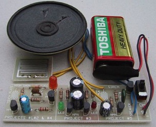 مدار داخلی واکی تاکی(اموزش ساخت)
 کتاب افزایش ممبر کانال تلگرام
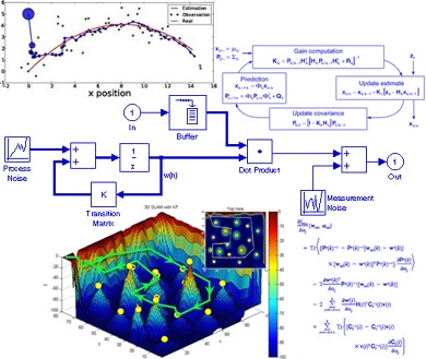 دانلود پاورپوینت فیلتر کالمن بر روی یک سنسور شتاب سنج برای تخمین سه متغیر حالت
 اندیکاتور ای کیو آپشن
 دانلود100% رایگان نرم افزار تبلیغات در تلگرام + آموزش کامل و فیلم آموزشی
moballegh1149@gmail.com


